In stock
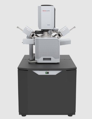
Field Emission - Scanning Electron Microscope (FE-SEM) Completed with Maps Mineralogy
Rp. 16.326.000.000
|
Type Brand Stock |
: : : |
Quattro S with Maps Mineralogy Thermo Scientific 2 Unit |
| Kategori | : | Alat Laboratorium |
| Jenis Produk | : | import |

BUSINESS PARK KEBON JERUK BLOK F 2 NO. 9, JL. MERUYA ILIR NO. 88, Desa/Kelurahan Meruya Utara, Kec. Kembangan, Kota Adm. Jakarta Barat, Provinsi DKI Jakarta, Kode Pos: 11620
sales@dynatech-int.com
Source :
- High-resolution field emission SEM column with a highstability Schottky field emission gun to provide stable high-resolution analytical currents
- 45° objective lens geometry with heated objective apertures
- Through-the-lens differential pumping reduces beam skirting for the most accurate analysis and highest resolution
Accelerating Voltage : Range 200 V to 30 kV
Probe Current : Beam current range: 1 pA to 200 nA
Resolution :
High Vacuum Imaging
- 1.0 nm @ 30 kV (SE)
- 2.5 nm @ 30 kV (BSE)
- 3.0 nm @ 1 kV (SE)
Low Vacuum Imaging
- 1.3 nm @ 30 kV (SE)
- 2.5 nm @ 30 kV (BSE)
- 3.0 nm at 3 kV (SE)
Extended vacuum mode (ESEM)
1. 1.3 nm at 30 kV (SE)
Magnification : 6 - 2.500.000x
Vacuum mode :
- High vacuum: < 6 x 10⁻⁴ Pa
- Low vacuum: up to 200 Pa
- ESEM: up to 4000 Pa
Vacuum system :
- 1 × 250 liter/s TMP
- 1 × PVP
- 2 × IGP
- Patented through-the-lens differential pumping
- Beam gas path length: 10 mm or 2 mm
Detector :
- ETD – Everhart-Thornley SE detector
- Low-vacuum SE detector (LVD)
- Gaseous SED (GSED) (used in ESEM mode)
- IR camera for viewing sample in chamber
Solid-State Detector Integration Kit :
- It allows having all three solid-state detectors connected simultaneously and provides support for up to twelve signal channels,
- This allows access to all solid-state detector segments
Retractable DBS Detector :
- Ultra-sensitive, solid state detector which is sensitive to emitted electrons from 500 V onward
- Mounted on a software-controlled retractable arm and allows simultaneous EDS spectra acquisition for WD ≥ 10 mm.
- An inner (materials contrast) and an outer (topography) concentric segment
- Detector can be used in high-vacuum, low-vacuum and in ESEM mode.
Dual EDS Detector :
- 2 unit of EDS Detector
- Silicon drift detector with active area of 30 mm2
- Detects elements from Be to Am
- Liquid nitrogen (LN2) free, using thermoelectric peltier cooling
- Mapping, LineScan, Spectrum acquisition
MAPS Mineralogy :
- Advanced fully automated spatial mineralogy solution combining ultra-high resolution imaging hardware with the highest throughput and anlytical performance to create accurate and statistically robust data
- Combines BSE imaging with EDS acquisition to build accurate pictures of particles and standard rock thin sections
- Advanced mineral classification approach combinies unique patent-protect capabilities including: Accurate, automated sub-micron identification and quantification of minerals, Direct mineral/mineral associations, Rapid multi-elemental mapping
- Advanced image analysis and reporting software
- Creation and viewing images, tables and charts
- General-purpose and proven application specific reports (Liberation, locking, grain size, etc.)
- Property based filtering, sorting and classification;
- Database management system to combine data from multiple batches, samples and size fractions into a single report;
- Built-in mineral database of mineral compositions, densities hardness and other properties;
- Export of images, tables, and charts to image analysis, word-processing and spread-sheet software;
- Over 4000 integrated mineral species;
- Stiching automation: Multi-scale acquisition, easy setup.
MAPS 3 :
- Maps provides automated acquisition of image mosaics via easy set up and offers complete control on location, resolution and imaging parameters,
- Maps corrects for non-linear stage behavior to increase navigation accuracy,
- Maps supports batch acquisition, allowing the user to schedule acquisition of multiple areas in one job, saving supervised time,
- Microscope real-time stitching of tiled images can be carried out concurrent with image acquisition,
- Maps also makes it easy to re-align and collect data over multiple imaging sessions
In-Chamber Nav-Cam :
- Automatic image acquisition with sample lighting
- 160 x 105 mm field of view
- 3072 x 2048 pixels or approximately 6 megapixels
- Digital zoom
- Image annotation
- Image save
Holder :
- Standard multi-sample SEM holder, uniquely mounts directly onto the stage, hosts up to 18 standard stubs (⌀ 12 mm)
- 10 x 30 mm block holder
- 16 x 25 mm block holder
Chamber :
- Inside width: 340 mm
- Analytical working distance: 10 mm
- Ports: 12
Sample Stage:
- Type : Eucentric goniometer stage,
- 5-axes motorized
- XY 110 × 110 mm
- Repeatability < 3.0 μm (@ 0° tilt)
- Motorized Z 65 mm
- Rotation n × 360°
- Tilt -15° / +90°
- Max. sample height Clearance 85 mm to eucentric point (10 mm)
- Max. sample weight 500 g in any stage position (up to 2 kg at 0° tilt)
- Max. sample size 122 mm diameter with full X,Y, rotation (larger samples possible with limited stage travel or rotation)
FESEM Image Processor :
- Dwell time range from 25 ns – 25 ms/pixel
- Up to 6144 × 4096 pixels
- File type: TIFF (8, 16, 24 bit), JPEG or BMP
- Single-frame or 4-view image display
- SmartSCAN™ (256-frame average or integration, line integration and averaging, interlaced scanning)
- DCFI (Drift Compensated Frame Integration)
System Control :
- 64-bit GUI with Windows, keyboard, optical mouse
- 24-inch LCD display, WUXGA 1920 × 1200
- Navigation montage Software
- Undo / Redo functionality
- Customizable graphical user interface, with up to 4 simultaneously active images
- User guidance software enabling new or infrequent users to obtain excellent results
Manual User Interface :
- Supplementary control console providing direct manual control of microscope parameters such as focus, magnification, contrast, brightness, beam shift and stigmator.
Maintenance Support : Remote Diagnostic
Compressor :
- Compressor 230 V, 50/60 Hz with 4-liter Tank
- Compressed air is required for operating pneumatic valves and the microscope's leveling system
- The compressor is connected to the mains supply unit of the microscope.
Chiller :
- Air-cooled Water Chiller 230 V, 50/60 Hz
- Continuous cooling between 5°C and 40°C
- Stability of 0.1 °C
- Cooling capacity: up to 1100/1300 W
Sample Standard :
- High resolution test specimen, Gold on Carbon, 1 pc
- Magnification calibration grating replica, 1 pc
- EDS calibration and contains ANSI 316 stainless steel, SiO2, Mn, B, C and a 200 mesh Cu grid in a 12.7mm aluminum pin stub
SEM Starter Kits :
- Polyprop Sorting Box,5 CMPT,EA, 1 pc
- Carbon Cond. Tape,12MM X 20M, 18 pcs
- Pin Mount AL.12.7X11MM,8MM PIN, 200 pcs
- SEM Pin MNT 25.4MM,9.5MM PIN H, 50 pcs
- Pelco 12.7MM Pin MNT Gripper, 1 pc
- Pelco 25MM Pin MNT Gripper, EA, 1 pc
- Storage Box For 14 Pin Mounts, 1 pc
- Split Specimen Mount, 1/2x1/8", 2 pcs
Materials for Installation :
- Uninterruptible Power Supply (UPS) GE LP 8 kVA
- 1 Unit of Gas Nitrogen UHP (99,9995%), 7,5 M3 (Includes cylinder, Hose, and regulators)
- EMO Box, Safety Emergency Off button for fast power shut downs with electronics
- Generator Set 10 kVA
- 1 Unit of Gas Argon, (Includes cylinder, Hose, and regulators)
- Working desk and chair for operator,
- Printer
- Installation dan Training
Sample Preparation :
- Sputter Coater with Au source
- Carbon Coater
- Precision low-speed saw
- Grinding and Polishing with Power Head completed with resin package
| Type | Quattro S with Maps Mineralogy |
| Merek | Thermo Scientific |
| Nama Alat | Field Emission - Scanning Electron Microscope (FE-SEM) Completed with Maps Mineralogy |
| Sub - Kategori | Alat Uji |
| Kategori | Alat Laboratorium |
Related Products
More Products
ISQ EM LIQUID CHROMATOGRAPHY MASS SPECTROMETRY LCMS SINGLE QUADRUPOLE
EVOLUTION ONE SPECTROPHOTOMETER UV-VIS
FLASHSMART CHN ORGANIC ELEMENTAL ANALZYER WITH MICROBALANCE
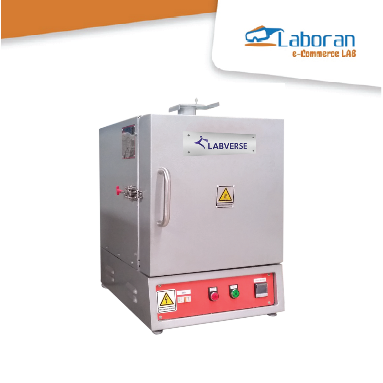
Labverse Furnace 1200 C
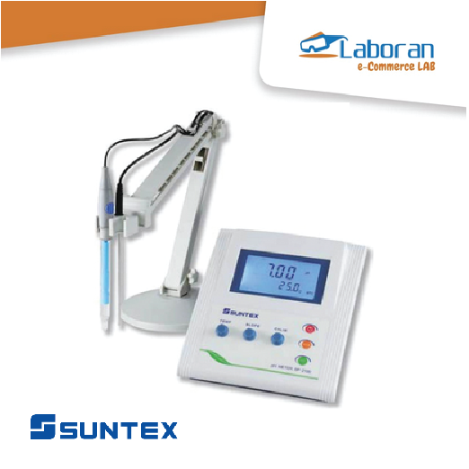
Suntex Laboratory pH/mV/TEMP Meter
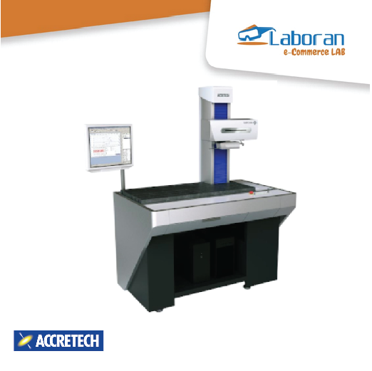
Accretech Hybrid Surface Roughness and Contour Tester
Suntex Conductivity Meter
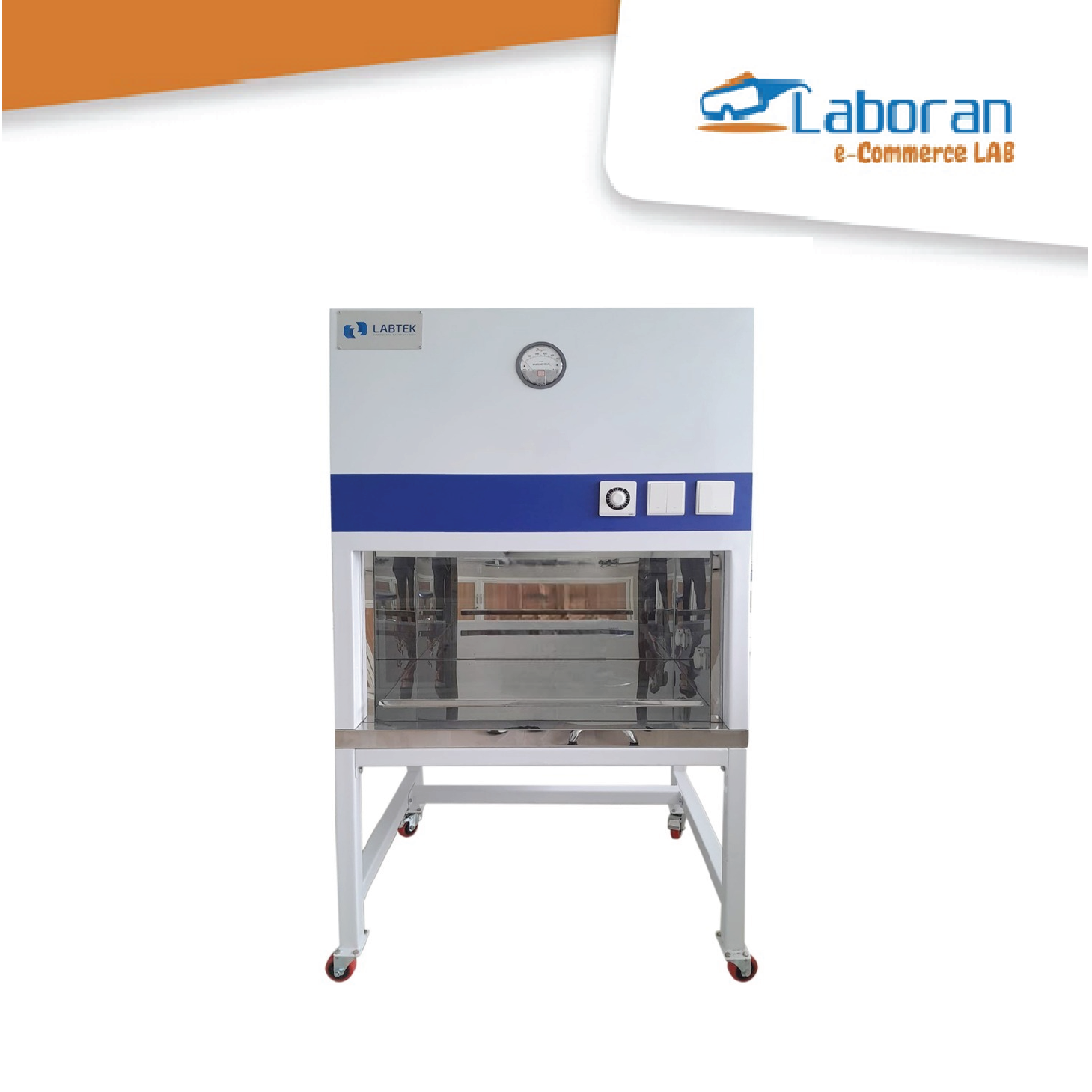
Air Flow Laminar
